Here is a selection of new images from our free, online textbook:
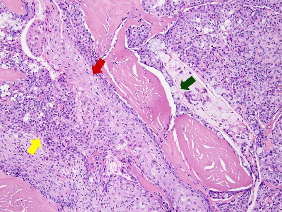
Salivary glands > Mucoepidermoid carcinoma
Solid areas of the tumor are formed by different proportions of epidermoid (squamous) cells (red arrow), mucus cells (green arrow) and intermediate cells (yellow arrow) (histopathology, H&E stain, 10x).
Contributed by Rema A. Rao, M.D. and Saeed Asiry, M.D.

Skin melanocytic tumor > Invasive melanoma
44 year old woman with 0.8 cm nodule on her back. Scanning magnification shows a nodular melanocytic proliferation with minimal junctional component (2x).
Contributed by Michele Donati, M.D.
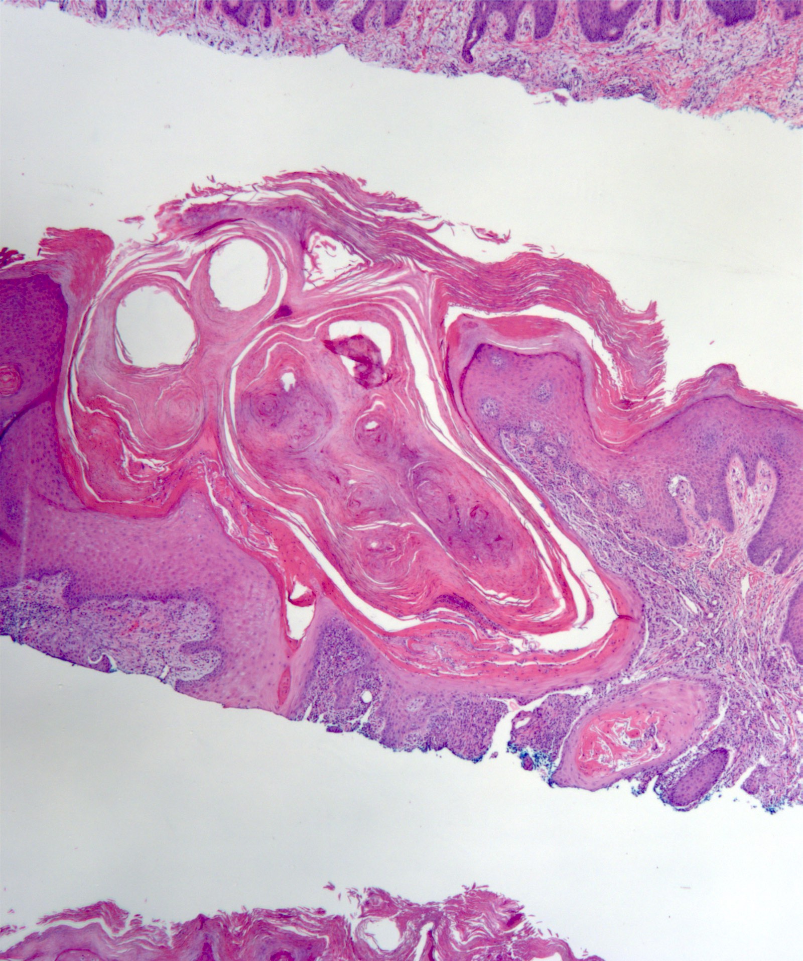
Skin nonmelanocytic tumor > Keratoacanthoma
An endophytic squamous proliferation with central crater filled with keratin.
Contributed by Poonam Sharma, M.B.B.S.

Bladder, ureter & renal pelvis > Urothelial carcinoma – invasive > Nested
Anastomosing, irregular, large nested and inverted growth of cytologically bland urothelium with desmoplastic stromal reaction (4x).
Contributed by Megan L. Brown, M.D. and Maria Tretiakova, M.D., Ph.D.
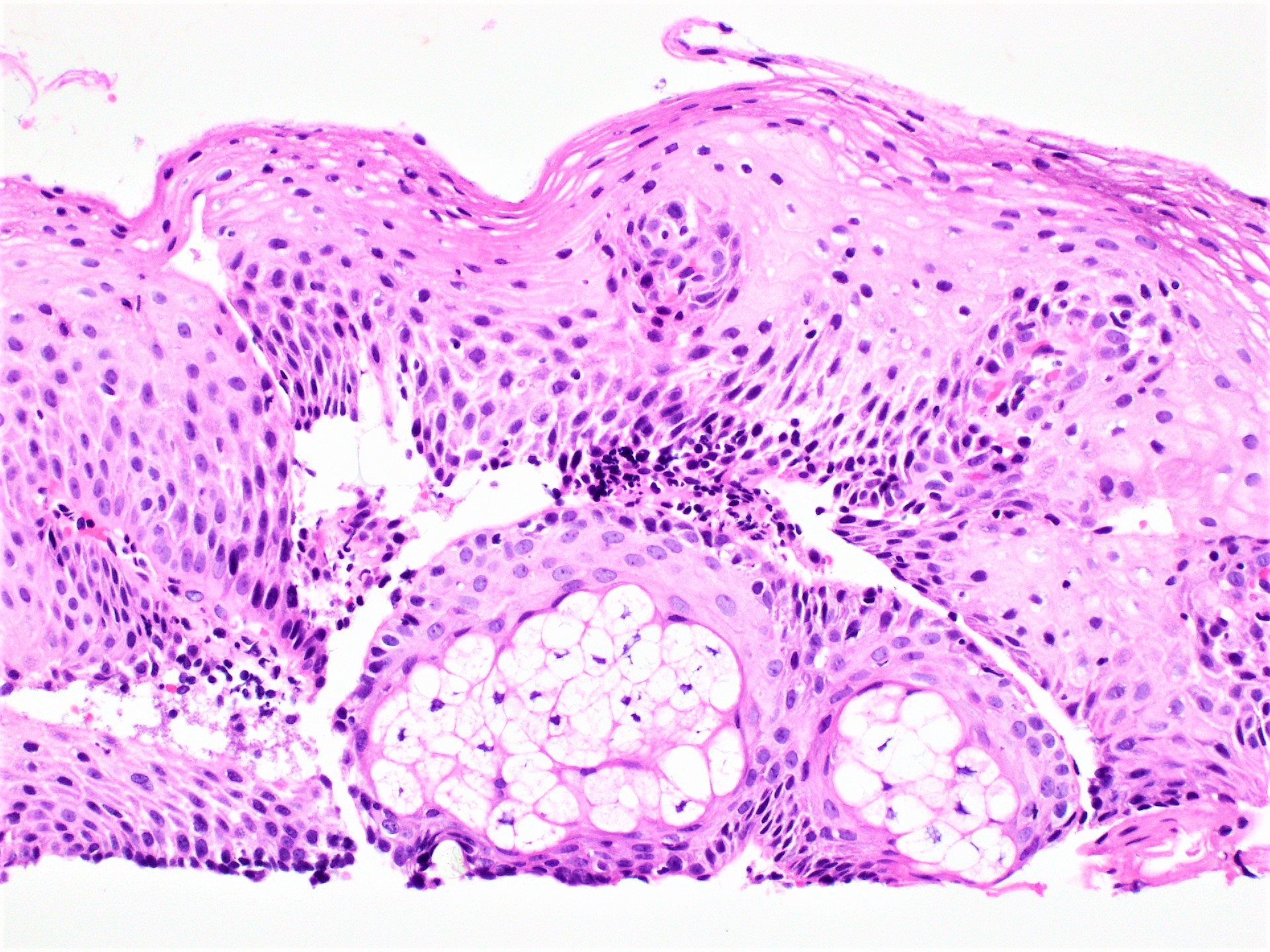
Esophagus > Ectopic sebaceous glands
Mature sebaceous gland within the lamina propria.
Contributed by Yukihiro Nakanishi M.D., Ph.D.
