29 November 2022: Image Quiz #96

Authors: Juwairiya Arshi, M.B.B.S., Aaron R. Huber, D.O.

Which histological feature is most common in celiac disease?

Select an answer:

A. Crypt hyperplasia

B. Granulomas

C. Intraepithelial lymphocytosis

D. Mucosal eosinophilia

E. Villous atrophy

To sign up for our biweekly image quiz newsletters and more, visit pathologyoutlines.com/subscribe.html.

4 August 2021: Pathology Images of the Week

Here is a selection of new images from our free, online textbook:

Lymphoma & related disorders > Prolymphocytic leukemia

Diffuse marrow involvement by medium to large sized cells with prominent nucleoli (200x, H&E).

Contributed by Richard K. Wood, M.D. and Dietrich Werner, M.D.

Ovary > Thecoma

Well circumscribed, firm, yellow-tan mass.

Contributed by Victoria Collins, M.D. and Tamara Kalir, M.D., Ph.D.

Prostate gland & seminal vesicles > Anatomy & histology

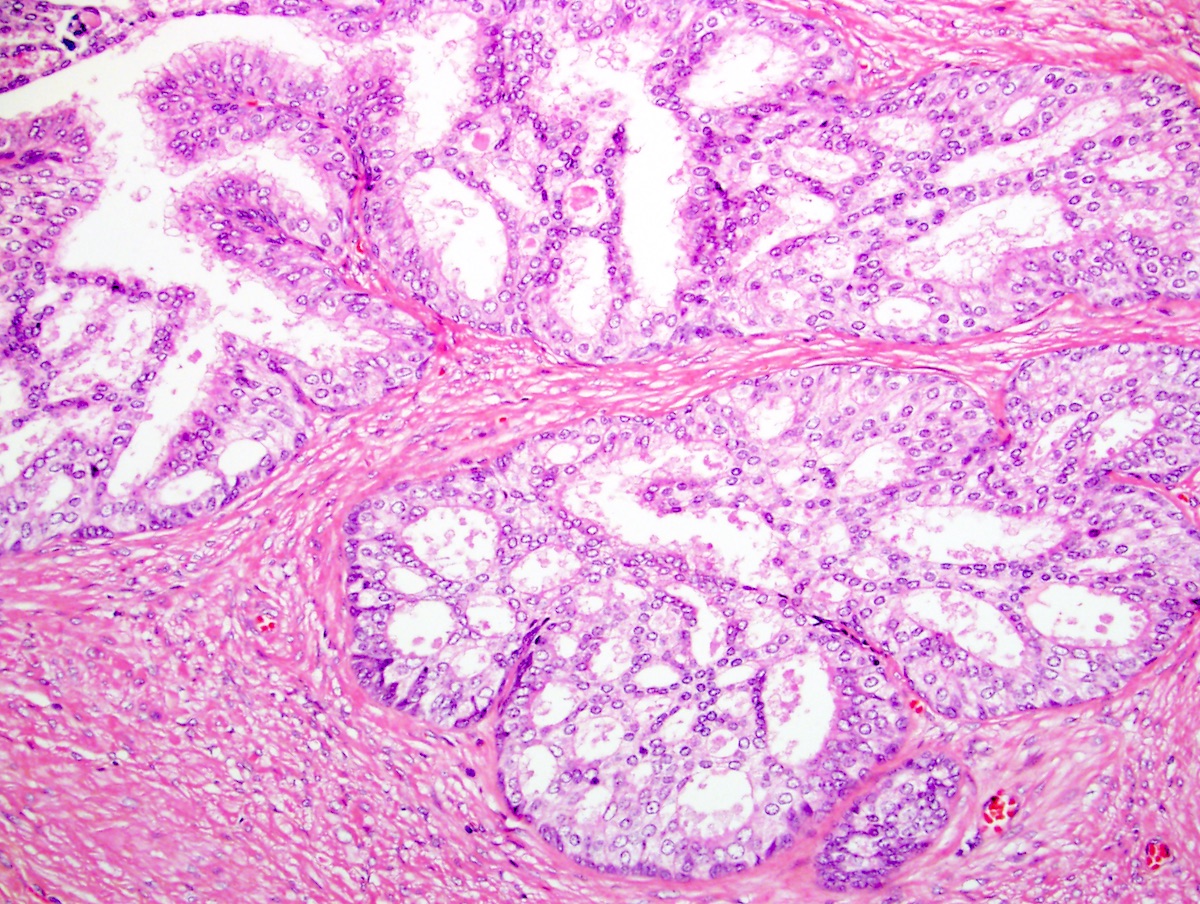
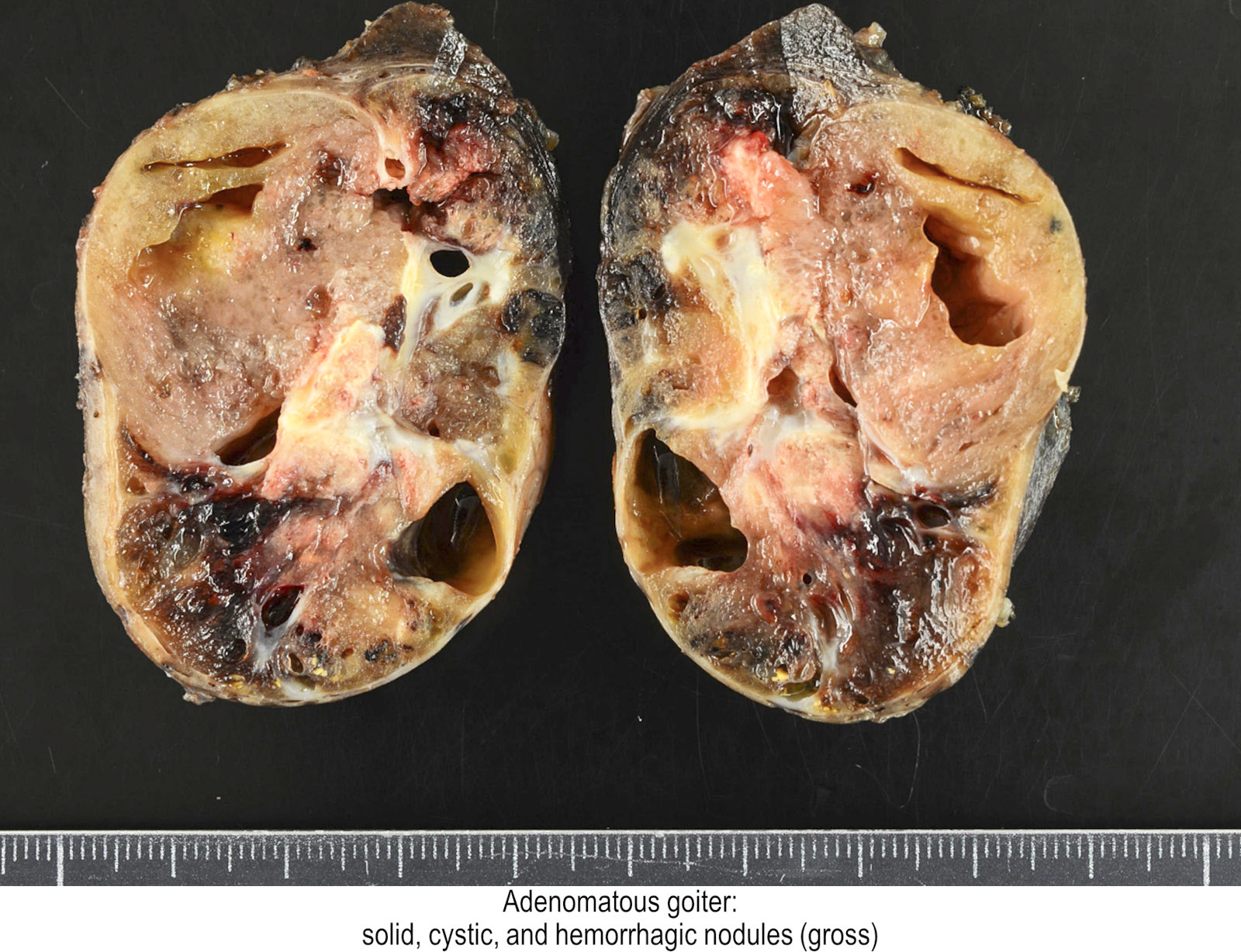
Here, the central zone cells are hyperplastic. Despite the cribriform structure, the bland nuclei rule out intraepithelial neoplasia or cribriform cancer. Also note the surrounding dense stroma of the central zone.

Contributed by Kenneth A. Iczkowski, M.D.

Prostate gland & seminal vesicles > Malakoplakia

Michaelis-Gutmann bodies highlighted by arrows.

Contributed by Theodorus van der Kwast, M.D., Ph.D.

Small intestine & ampulla > GIST

Small intestinal GIST with skeinoid fibers.

Contributed by Shefali Chopra, M.D.

7 July 2021: Pathology Images of the Week

Here is a selection of new images from our free, online textbook:

CNS tumor > Papillary craniopharyngioma

Classic low power cauliflower-like configuration with smooth borders.

Contributed by Nelli S. Lakis M.D., M.Sc.

Nasal cavity, paranasal sinuses, nasopharynx > Sinonasal undifferentiated carcinoma

Sheets of undifferentiated malignant epithelial cells with vesicular nuclear chromatin and prominent nucleoli.

Contributed by Bin Xu, M.D., Ph.D. and Abeer Salama, M.D.

Placenta > Choriocarcinoma

Intraplacental choriocarcinoma showing atypical syncytiotrophoblast, cytotrophoblast and intermediate trophoblast adjacent to uninvolved chorionic villi (H&E, 200x).

Contributed by Tatyana Zinger, D.O.

Small intestine & ampulla > Heterotopic gastric mucosa

Polypoid lesion predominantly lined by gastric foveolar epithelium and small intestinal epithelium on the left side.

Contributed by Khalid Amin, M.D.

Liver & intrahepatic bile ducts > Hepatocellular adenoma

HNF1A mutated hepatocellular adenoma: low power view showing well defined border between the lesion (right) and background liver (left).

Contributed by Monica Abdelmalak, M.D.

17 March 2021: Images of the Week

Here is a selection of new images from our free, online textbook:

Skin nontumor > Warts (verrucae)

Hyperkeratosis, papillomatosis, hypergranulosis, koilocytosis and inward bending of rete ridges at borders of lesion. Dermal papillae show dilated capillaries.

Contributed by Heba Ahmed Abdelkader, M.D.

Small intestine & ampulla > Lymphangioma

Scanning magnification shows the dilated lymphatics in the submucosa of the small intestine (H&E, 2x).

Contributed by Haider A. Mejbel, M.D.

Stomach > Pyloric gland adenoma

High power view of PGA showing small tubular glands with low cuboidal epithelium and bland nuclei.

Contributed by Supriya Srivastava M.D., Ph.D.

Bone & joints > Adamantinoma

Anastomosing thin trabeculae of mildly atypical epithelioid and spindled cells within fibrous stroma.

Contributed by Borislav A. Alexiev, M.D.


How can you search the textbook?

For desktop / tablet, you can either use the Google search bar in the upper right corner or visit PathologyOutlines.com and then scroll down to where the 60 chapters are listed, as demonstrated below.

For mobile, visit PathologyOutlines.com, click on “Chapters by Subspecialty” (as demonstrated below) and then click on the subspecialty, chapter and topic.

For any device you can use your favorite browser and type in PathologyOutlines.com in addition to the search term.

3 March 2021: Images of the Week

Here is a selection of new images from our free, online textbook:

Cervix > HPV related adenocarcinoma (usual type and variants)

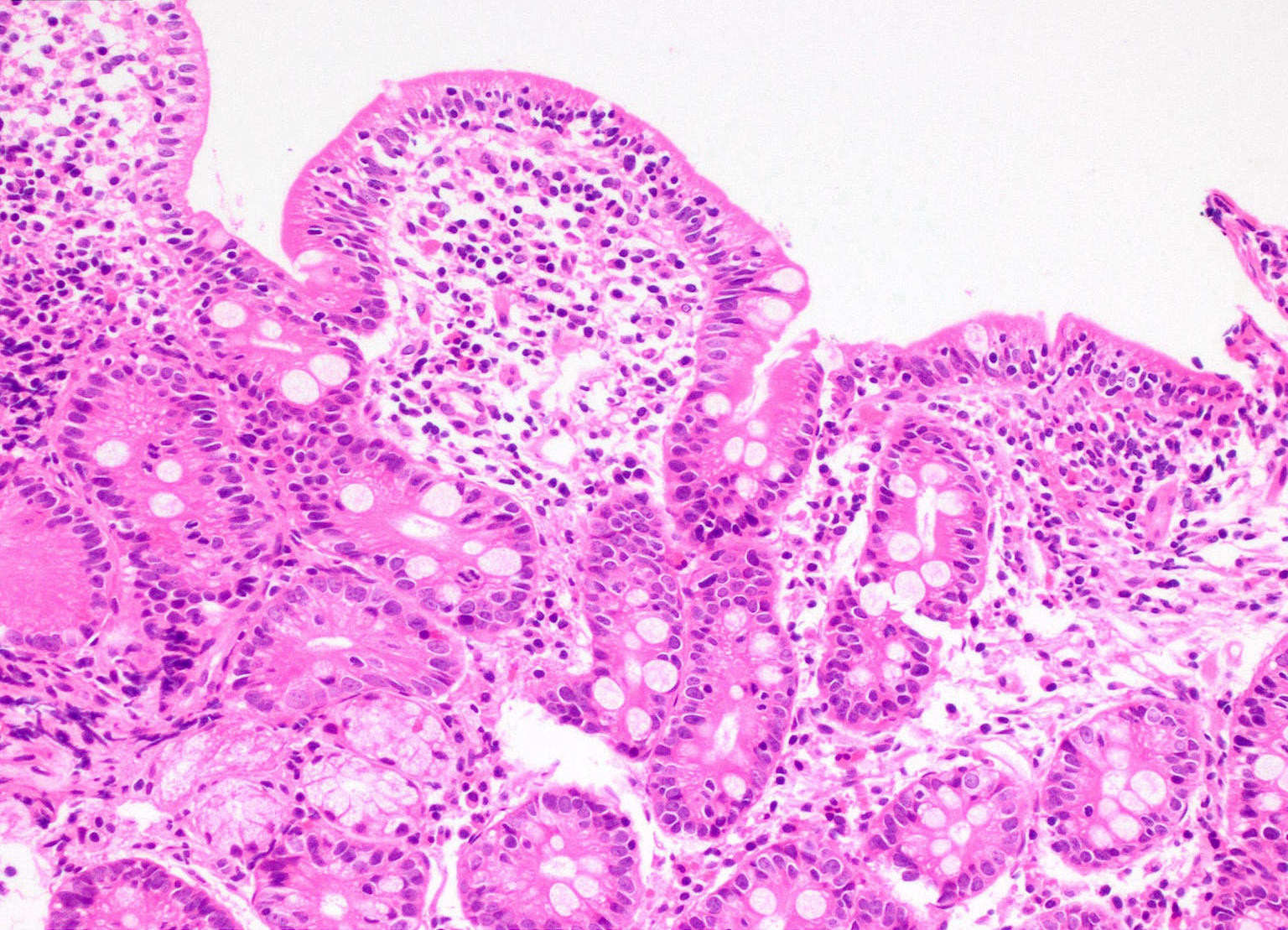
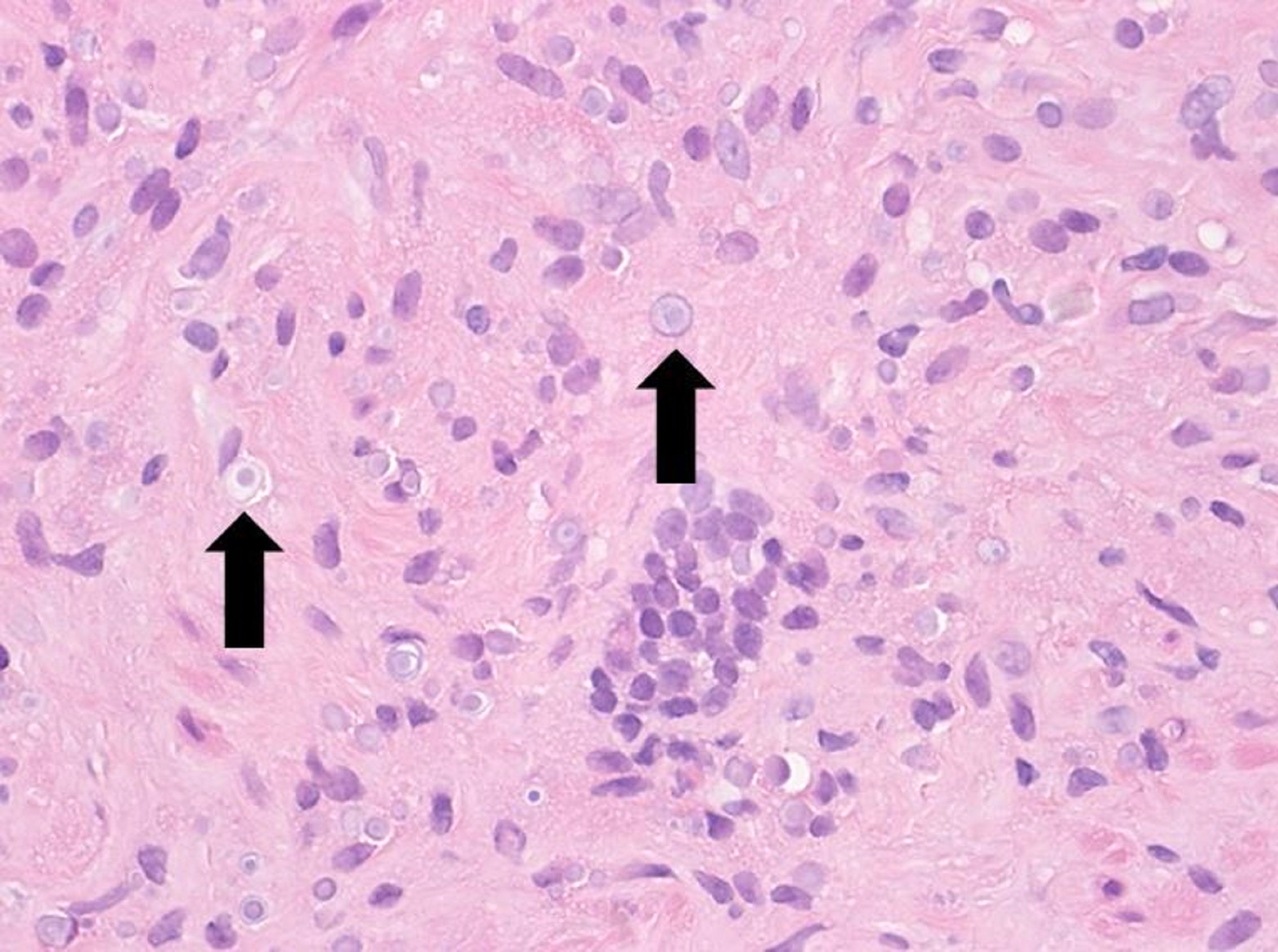
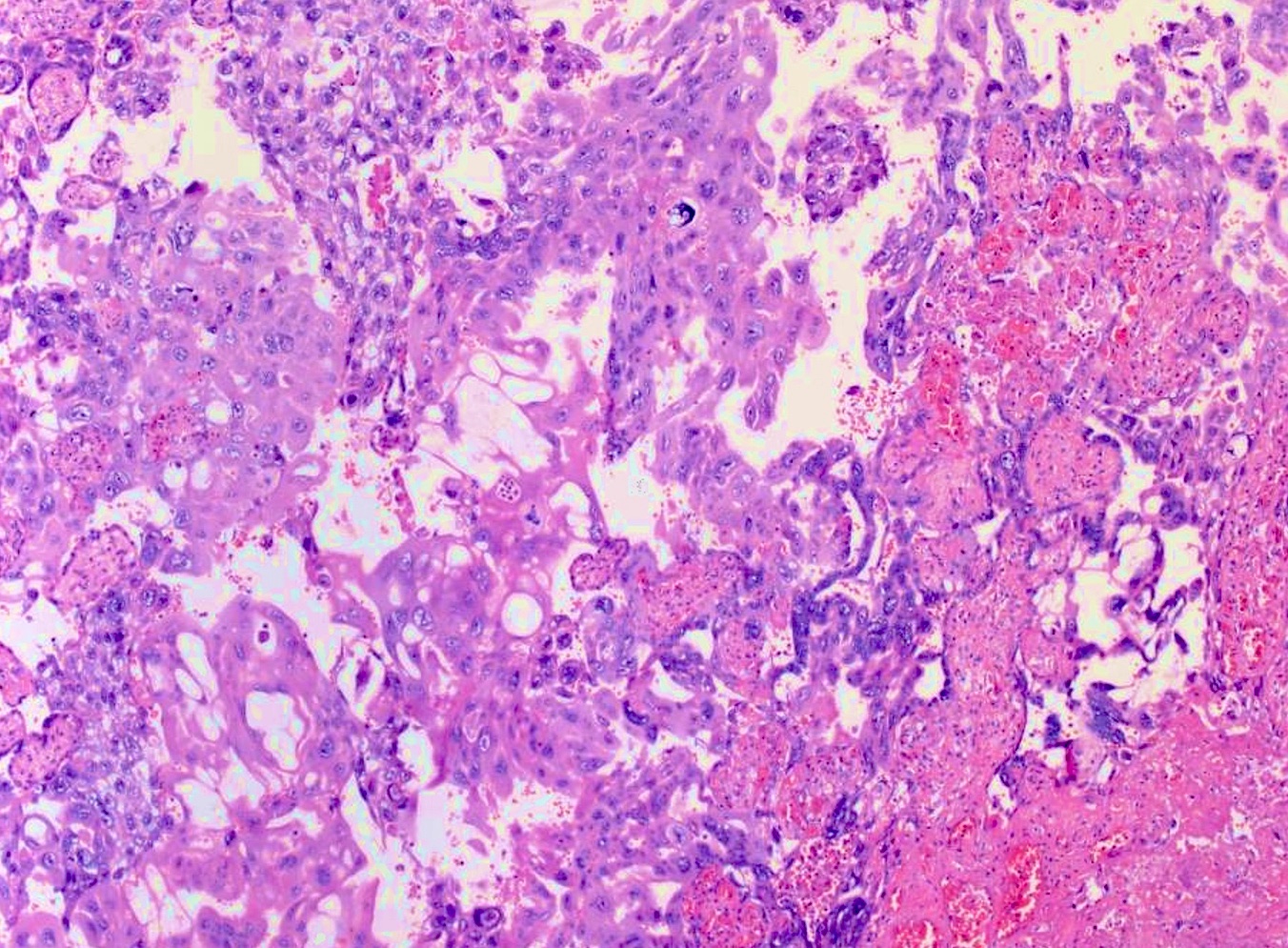
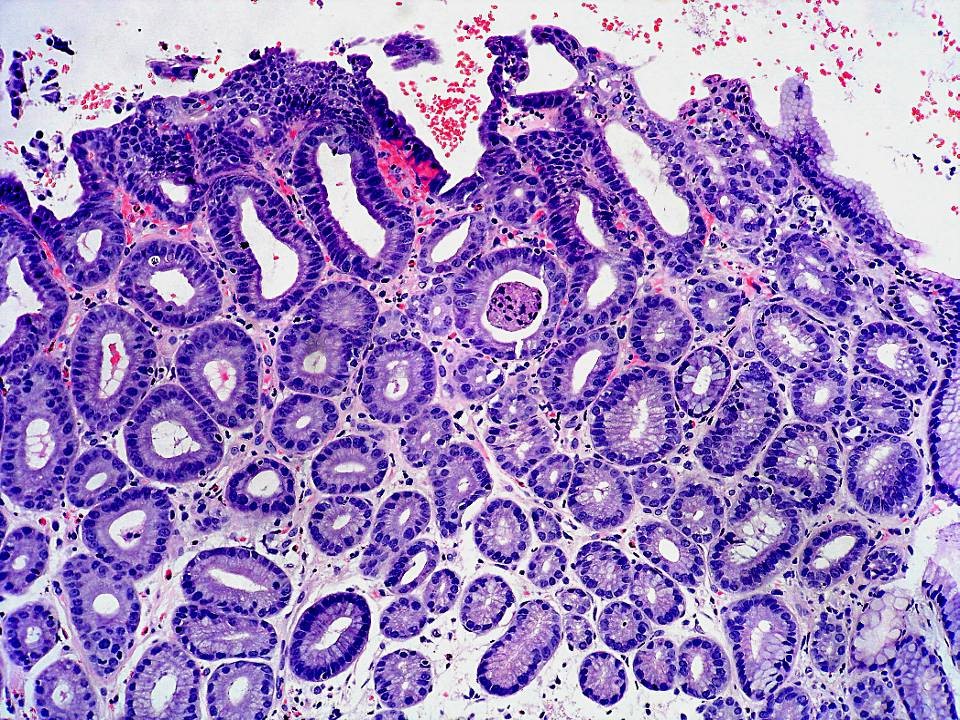
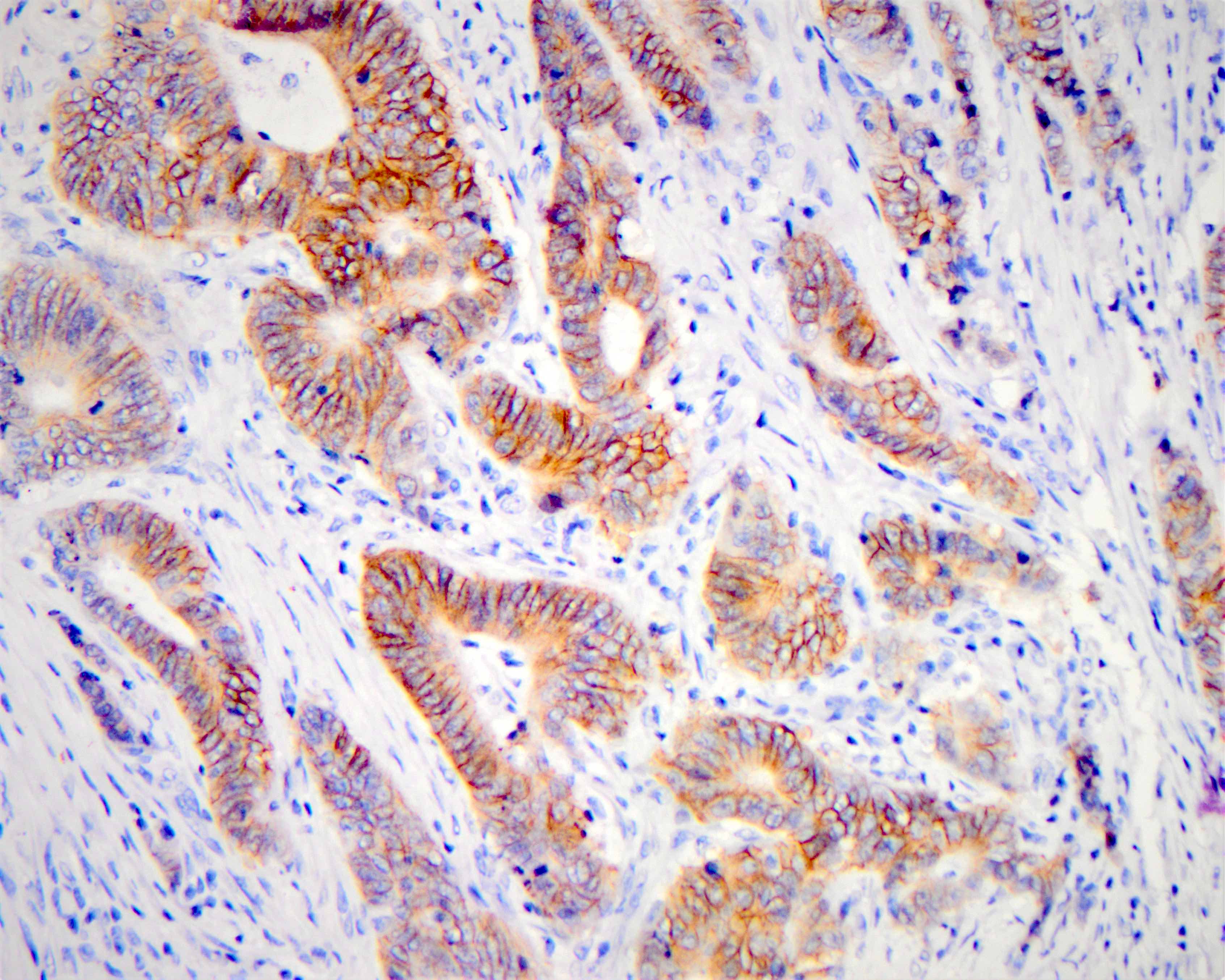
Invasive adenocarcinoma, pattern B (H&E stain). Early destructive invasion, in an otherwise well differentiated (pattern A) neoplasm is seen as small irregular glands with prominent inflammation and stromal desmoplasia.

Contributed by Carlos Parra-Herran, M.D.

Stains & molecular markers > HER2 colon

Score 3+, HER2 IHC: the tumor cells of this colorectal adenocarcinoma show strong circumferential membrane staining (200x).

Contributed by Roula Katerji, M.D. and Aaron R. Huber, D.O.

Stains & molecular markers > Pan-TRK (EPR17341) [NTRK]

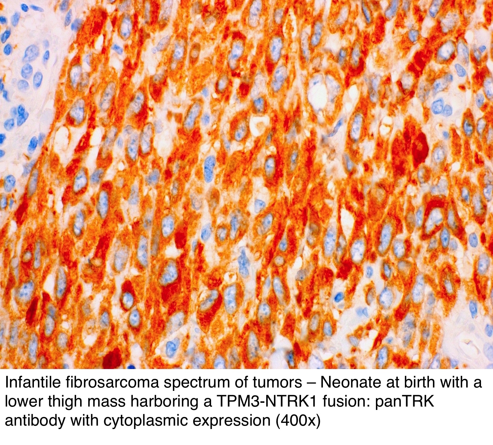
Infantile fibrosarcoma spectrum of tumors – neonate at birth with a lower thigh mass harboring a TPM3-NTRK1 fusion: panTRK antibody with cytoplasmic expression (400x).

Contributed by Jessica L. Davis, M.D.

Liver & intrahepatic bile ducts > Hepatocellular carcinoma overview

Hepatectomy for hepatocellular carcinoma demonstrating large cytoplasmic hyaline eosinophilic inclusions and trabecular architecture (400x).

Contributed by Irene Y. Chen, M.D.

Small intestine & ampulla > Adenocarcinoma-small intestine

Diagnostic low magnification frozen section image showing complex glandular architecture and desmoplastic stromal reaction (H&E, 40x).

Contributed by Krutika S. Patel, M.B.B.S., M.D. and Annika L. Windon, M.D.


How can you search the textbook?

For desktop / tablet, you can either use the Google search bar in the upper right corner or visit PathologyOutlines.com and then scroll down to where the 60 chapters are listed, as demonstrated below.

For mobile, visit PathologyOutlines.com, click on “Chapters by Subspecialty” (as demonstrated below) and then click on the subspecialty, chapter and topic.

For any device you can use your favorite browser and type in PathologyOutlines.com in addition to the search term.